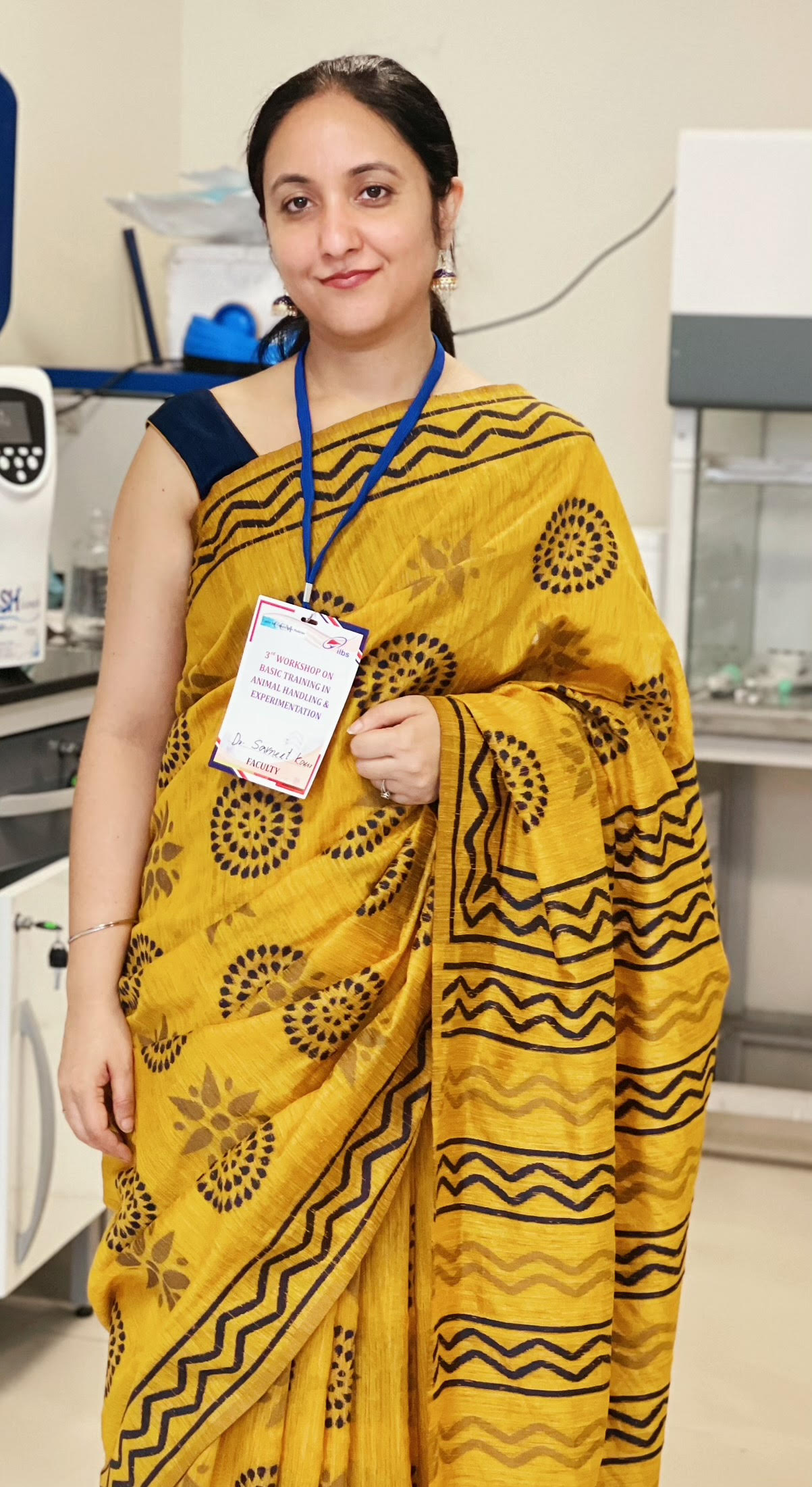
In Relationship: Liver with Heart

By Dr. Savneet Kaur

Sitting in the upper right-hand side of the abdominal cavity, beneath the diaphragm, is the largest internal organ of our body, ‘the liver.’ Despite being involved in more than 500 vital functions, including metabolism, energy homeostasis, bile synthesis, detoxification, and synthesis, it often remains an overlooked and neglected organ. In this 3-part scientific series, you will learn about how the liver connects with other organs, why liver health is essential for our overall well-being, and how to maintain our liver by observing a healthy lifestyle.

Part 2 - Take care of your liver for a healthy heart!​

The heart and the liver share a close rapport with each other and an ailment in one affects the other. Many patients with liver disease, irrespective of the etiology, show a diverse array of cardiovascular (CVD) abnormalities such as systolic and diastolic heart failure, myocardial fibrosis, and inflammation without any prior cardiac disease (1). Here we learn some interesting physiology concepts of how liver disease can foster CVD abnormalities. 

To have deeper insights into how the liver affects the heart, we need to first understand the schema of blood circulation in the liver. The liver receives blood from two sources, the portal vein (75% of total blood supply) and hepatic artery (25% of total). The portal vein collects the substrate-rich venous blood from the spleen and all organs participating in the digestion of food, such as the stomach, pancreas, small bowel, and the colon and then delivers it to the liver while the hepatic artery brings oxygen-rich blood from the heart. Liver either filters and processes all useful and non-useful products before either releasing them back into the hepatic veins and subsequently into the heart via the inferior vena cava (IVC) or stores them for later use.

During chronic and progressive liver injury (due to any etiology like viral infection, drugs, fat deposition, alcohol injury etc), there is increased cell death and deposition of scar tissue or fibrosis in the liver that causes structural alterations in the blood vessels inside the liver. These changes in the blood vessels result in a resistance to the blood flow that enters the liver from the portal vein, subsequently causing an increase in portal vein pressure, termed as portal hypertension. Normal value of portal pressure is 5 to 10 mmHg, which exceeds IVC pressure by 4 to 5 mmHg (portal/hepatic venous pressure gradient). A hepatic venous pressure gradient (HVPG) >5 mmHg defines portal hypertension. An enhanced HVPG makes the portal blood to bypass the liver and re-routes it through portal-systemic collateral vessels which are newly formed abnormal veins in the intestine and stomach that connect portal veins directly to IVC and heart. These collaterals induce an increase in venous return to the heart. Prolonged and severe portal hypertension, higher are the number of portal-systemic collateral vessels (2).

An overview of relationships between portal hypertension, splanchnic vasodilation, hyperdynamic circulation, systemic effects and heart during early and advanced stages of liver disease. BP: Blood Pressure; RAAS: Renin-Angiotensin-Aldosterone System; SNS: Sympathetic nervous system.

A direct link between the gut and the heart detouring the liver leads to deleterious consequences. The opening of new portal-systemic collaterals participates in a further increase in the portal blood inflow due to dilation of the splanchnic/abdominal and systemic blood vessels and hence a decreased systemic vascular resistance. The dilation happens due to an increased release of several intestinal vasoactive and vasodilatory factors including nitric oxide, prostacyclin, carbon monoxide by the splanchnic and systemic blood vessels and also due to the splanchnic vascular hypo-responsiveness to vasoconstrictors. This state of systemic vasodilation, low vascular resistance and a fall in systemic arterial blood pressure leads to a sequelae of events comprising of activation of vasoconstriction systems in the kidney like neurotransmitters and neurohormonal mediators including norepinephrine, renin–angiotensin–aldosterone system (RAAS) and vasopressin resulting in a compensatory rise in the cardiac output and heart rate which is termed as a state of hyperdynamic circulation. RAAS is a complex multi-organ hormonal system that is activated whenever there is a drop in the blood pressure. It increases water and electrolyte reabsorption in the kidneys and thus aids to elevate systemic blood volume and pressure. An activation of the RAAS system augments hyperdynamic circulation and cardiac output which puts extra burden on the heart to pump out more blood, leading to the development of cardiac dysfunction over time. As the liver disease further worsens, the decrease in systemic vascular resistance becomes so intense that the cardiac output fails to increase further, thus ultimately leading to a high-output heart failure. With increase in the severity of liver disease, splanchnic and systemic vasodilation and low arterial blood pressure, there is a reduction in the arterial blood flow to vascular beds of other organs like kidney and lungs as well. Thus, hyperdynamic circulatory syndrome is the pathogenetic basis not only for heart ailments and failure but also for the development of several other complications including hepato-renal syndrome, hepato-pulmonary syndrome, shock susceptibility and tissue hypoxemia (2). Besides vasoactive substances, several other toxins, metabolic products from the injured gut, that are normally detoxified in the liver now enter the IVC and systemic blood, inflicting damage to the blood vessels of the lung and the heart leading to aberrations. Also, many inflammatory and cardio-suppressive factors are derived directly from the diseased liver itself into the blood circulation. These systemic inflammatory factors contribute to the development of atherosclerotic lesions and several other abnormalities in the heart by mediating biochemical, structural and functional changes in the heart cells or cardiomyocytes. About 50% of patients with end-stage liver disease or cirrhosis show changes in the heart collectively called ‘cirrhotic cardiomyopathy’ (3). To explore the relationship between the severity of liver disease and the degree of myocardial involvement, a recent study used both cardiac and liver imaging in patients with liver cirrhosis. Compared with control participants, the subjects with liver cirrhosis displayed reduced myocardial functions and elevated markers of cardiac disease. Interestingly, liver stiffness (an indicator of liver fibrosis) emerged as an independent predictor for myocardial fibrosis (4).

In another study, it was shown that not only end-stage liver cirrhosis patients but also those with a higher amount of liver fat show early signs of ‘hardening of arteries’ or atherosclerosis and have a 1.5 fold higher risk of coronary microvascular dysfunction (inappropriate blood flow in small blood vessels of the heart) and hence increased CVD risk as compared with those who did not have liver fat (5). Increased amount of fat in the liver causes a disease called non-alcoholic fatty liver disease (NAFLD). NAFLD is a hepatic manifestation of metabolic syndrome with a prevalence of 38.6% in the adult population in India. In subjects with obesity and type 2 diabetes, its prevalence is as high as 65-90% and represents the most common cause of chronic liver disease and liver transplantation worldwide. NAFLD encompasses a spectrum of diseases starting from steatosis (liver fat accumulation >5% of liver weight) to non-alcoholic steatohepatitis or NASH (with liver fat, cell damage and inflammation) to NASH with fibrosis and cirrhosis. Patients with NASH have a higher cell damage and portal blood flow disturbances and are hence more prone to develop heart ailments than patients with simple steatosis. Long-term follow-up studies demonstrate CVD mortality to be the most important cause of death in patients with NASH.
 
The mechanisms by which a fatty liver might contribute to CVD are complex and just begun to be understood. A liver full of fat might amplify fat deposition in the blood vessels and the heart, leading to atherogenesis and myocardial steatosis. Atherogenesis, we all know, causes stiffening of the aorta, affecting myocardial function by increasing left ventricular afterload. This means that the left ventricle of the heart must now work more to eject the same amount of blood into the aorta during systole, leading to thickening of its wall and an increase in its oxygen requirements, ultimately predisposing to heart failure (6). Increased cardiac output and hyperdynamic circulation due to changes in intrahepatic blood vessels in patients with NASH may represent an additional risk factor for CVD. Altered blood levels of various endothelial and coagulation proteins (that affect inflammation, bleeding and blood clot formation) such as von Willebrand factor, plasminogen activator inhibitor, vascular endothelial growth factor, factor VIII, IX, XI and XII, is also one of the key reasons for development of atherosclerotic CVD. Patients with NASH and other liver diseases show a higher procoagulant imbalance than those with no liver disease. The two major naturally occurring anticoagulants (antithrombin and protein C) are known to be decreased in NASH and cirrhosis while Factor VIII (one of the most powerful procoagulants) has been demonstrated to be on the higher side in these patients (7). This imbalance might play a role in the risk of CVD events by affecting the process of thrombosis or formation of blood clots in the vessels. Thrombosis or blood clots constitute a crucial step in blocking of the coronary arteries and hence the pathogenesis of ischemic heart disease.
 
CVD abnormalities in early stage liver disease patients are usually clinically silent or a mild entity and hence most often ignored. However, all patients with liver anomalies should closely monitor their heart and regularly perform CVD risk assessment. Although, we do not have any specific treatments yet for NAFLD and NASH except for lifestyle modifications including exercise and a healthy diet, yet a regular follow-up of heart would allow the identification of high-risk patients who are candidates for adequate therapeutic interventions for cardiac ailments. Heart abnormalities also affect the patient’s prognosis during and after procedures such as surgery and liver transplantation. A vigilant cardiac evaluation is therefore pertinent prior and post to these surgical procedures as well.
 
Wondering how a diseased heart affects a healthy liver? Well this is a discrete entity termed as ‘Cardiac hepatopathy’ and thus needs a separate deliberation.

References

  1. Fede G, Privitera G, Tomaselli T, Spadaro L, Purrello F. Cardiovascular dysfunction in patients with liver cirrhosis. Ann Gastroenterol. 2015;28(1):31-40.
  2. Møller S, Bendtsen F. The pathophysiology of arterial vasodilatation and hyperdynamic circulation in cirrhosis. Liver Int. 2018;38(4):570-580. 
  3. Møller S, Lee SS. Cirrhotic cardiomyopathy. J Hepatol. 2018 Oct;69(4):958-960. 
  4. Isaak A, Praktiknjo M, Jansen C, Faron A, Sprinkart AM, Pieper CC, Chang J, Fimmers R, Meyer C, Dabir D, Thomas D, Trebicka J, Attenberger U, Kuetting D, Luetkens JA. Myocardial Fibrosis and Inflammation in Liver Cirrhosis: MRI Study of the Liver-Heart Axis. Radiology. 2020;297(1):51-61.
  5. Vita T, Murphy DJ, Osborne MT, Bajaj NS, Keraliya A, Jacob S, Diaz Martinez AJ, Nodoushani A, Bravo P, Hainer J, Bibbo CF, Steigner ML, Taqueti VR, Skali H, Blankstein R, Di Carli MF, Dorbala S. Association between Nonalcoholic Fatty Liver Disease at CT and Coronary Microvascular Dysfunction at Myocardial Perfusion PET/CT. Radiology. 2019;291(2):330-337. 
  6. Kasper P, Martin A, Lang S, Kütting F, Goeser T, Demir M, Steffen HM. NAFLD and cardiovascular diseases: a clinical review. Clin Res Cardiol. 2021;110(7):921-937. 
  7. Tripodi A, Fracanzani AL, Chantarangkul V, Primignani M, Fargion S. Procoagulant imbalance in patients with non-alcoholic fatty liver disease. J Hepatol. 2017;66(1):248-250. 

About author

Dr. Savneet Kaur pursued Ph.D. from IGIB, Delhi, and DBT-postdoctoral fellowship from SCTIMST, Kerala. Currently, she is an Assistant Professor in the ‘Institute of Liver and Biliary Sciences (ILBS),’ New Delhi. Her research in ILBS revolves around vascular biology of the liver, deciphering cellular and molecular mechanisms underlying liver inflammation and fibrosis. She has significantly contributed to understanding how bone marrow-derived endothelial cells interact with resident liver cells and participate in liver fibrosis. For her work, she has received the prestigious young investigator award from the ‘American Association for the Study of Liver Disease.’ Current efforts of her team are directed towards developing endothelial cell-based targeted therapies for liver diseases. Dr. Savneet is a nominated member of the CPCSEA, Govt of India, and serves as ‘Scientist-in-charge’ of the Animal House Facility in ILBS. Besides the lab, she likes experimenting and innovating in her kitchen.

Dr. Savneet Kaur

Edited by: Dolly Singh