Research Summary: Developing an intuitive platform for radiomics and radiogenomics analysis that can aid in guiding personalized planning for glioma patients.
Author Interview
Kavita Kundal is a researcher exploring radiomics and radiogenomics, focusing on identification of non-invasive prognostic markers in glioblastoma to improve personalized patient care.
Linkedin: https://www.linkedin.com/in/kavitakundal
Twitter: https://x.com/kundalkavita
Lab: Dr. Rahul Kumar, Indian Institute of Technology Hyderabad (IITH)
Lab website: https://people.iith.ac.in/rahulk/
What was the core problem you aimed to solve with this research?
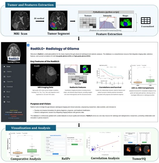
Glioma, a brain tumor arising from glial cells, is one of the most complex and heterogeneous cancers of the central nervous system. While Magnetic Resonance Imaging (MRI) is the standard first-line tool for detecting brain tumors, it primarily provides anatomical and structural information. To understand tumor biology, aggressiveness, and genetic alterations, clinicians still rely on biopsies, where a small tissue sample is extracted for pathological and molecular testing. However, biopsies are invasive, risky especially in sensitive brain regions and capture only a small portion of the tumor, often failing to represent its full spatial and biological heterogeneity. Gliomas frequently exhibit diverse molecular signatures across different regions, meaning a single biopsy may miss critical information. Considering the brain’s delicate nature, obtaining comprehensive tumor information invasively is both challenging and potentially dangerous. This creates a clear need for non-invasive approaches capable of extracting comparable biological insights from MRI scans. Our goal was to develop an imaging-based alternative that provides molecular-level information similar to biopsies. We focused on radiomics, which extracts quantitative features from medical images, to identify imaging biomarkers that reflect tumor biology and correlate with genetic and clinical outcomes.
How did you go about solving this problem?
We approached this problem through a combination of computational modeling, data integration, and platform development. First, we extracted thousands of quantitative radiomic features from MRI scans that describe the tumor’s shape, texture, intensity, and spatial variation. These features capture subtle patterns that are invisible to the human eye but can reveal underlying tumor heterogeneity and behavior. Using multi-institutional datasets that included MRI scans, survival information, and genetic data, we built a survival model to identify which helps in patient stratification into high- and low-risk categories. This analysis led to the creation of RaSPr (Radiomic Survival Predictor), an in-house model based solely on MRI-derived data. To make these analytical capabilities accessible, we developed a comprehensive and interactive web-based platform named RadGLO (Radiology of Glioma). RadGLO integrates radiomics, radiogenomics, and in-house developed models into a unified interface. It includes RaSPr for survival risk prediction and another developed module called TumorVQ (Tumor Volume Quantifier), which enables region-specific tumor volume estimation using MRI scans. Users can upload their MRI images, segmented tumor regions, or extracted feature files to perform analyses directly through the platform. Additionally, RadGLO includes a DataViz feature where users can explore each radiomic feature, perform radiogenomic analysis through correlation analysis module, which allows users to explore associations between over 20,000 genes and radiomic features. This helps identify imaging biomarkers that mimic genetic functions and contribute to understanding tumor progression and molecular mechanisms. The platform’s goal was not only to identify non-invasive markers but also to democratize access to advanced radiomic analysis by providing an intuitive, research-oriented tool for clinicians, students, and scientists worldwide.
This research work underlines the importance of the emerging field of radiogenomics in identifying non-invasive biomarkers of glioblastoma. — Dr. Rahul Kumar
How would you explain your research outcomes (Key findings) to the non-scientific community?
In simple terms, our research shows that MRI scans can reveal far more information than what is visible to the human eye. By using advanced computational methods, we can extract meaningful patterns called Radiomic Features from these images that reflect how aggressive a tumor is, how it might respond to treatment, and how long a patient might survive.
We built a web-based tool, RadGLO, that allows anyone to upload brain tumor MRI scans and automatically obtain predictions about tumor risk, grade, and even potential links with genetic changes. This makes it possible to gain deep biological insights from simple MRI images without requiring invasive biopsies. In essence, our work turns standard MRI scans into a non-invasive digital biopsy, enabling doctors and researchers to understand tumors more comprehensively and plan treatments more precisely.
What are the potential implications of your findings for the field and society?
The implications of this work are both scientific and societal. Scientifically, RadGLO demonstrates how imaging and genomics can converge to create new pathways for precision medicine. It establishes a foundation for integrative radiomics, where quantitative image analysis and molecular data are analyzed together to improve diagnostic accuracy, predict treatment response, and support clinical decision-making. From a societal perspective, this platform has the potential to reduce the dependency on invasive biopsies, minimizing patient risk, and discomfort. RadGLO’s open and user-friendly framework also encourages collaboration and data sharing within the research community. It supports the development of explainable AI models that clinicians can trust and interpret, bringing computational precision closer to real-world medical practice.
What was the exciting moment during your research?
The research domain itself is very exciting, every day we learn something new and see how science can explain phenomena that seemed mysterious before. For me, the truly exciting moment began with the topic itself. In India, studies on radiomics and radiogenomics were not widely explored when I first learned about them. I was fascinated by the idea that, without inserting a needle into the body, we could gain deep insights into tumor characteristics just by analyzing imaging that almost everyone undergoes at the hospital. This concept of extracting molecular and biological information non-invasively was both inspiring and motivating. From that moment, I dedicated myself to building expertise in the field, exploring advanced computational techniques, and developing tools like RaSPr, TumorVQ, and RadGLO. The best moment came when we saw that methods in radiomics could predict patient survival and reveal important details about tumor biology using only MRI scans. Knowing that advancements in this field could one day help reduce the need for risky biopsies and improve patient care made the experience truly rewarding.
Paper reference: Kundal, K., Rani, K.D., D, V. et al. RadGLO: an interactive platform for radiomic feature analysis and prognostic modeling in glioma. npj Precis. Onc. 9, 323 (2025). https://doi.org/10.1038/s41698-025-01124-z
Webserver: https://project.iith.ac.in/cgntlab/radglo/
Explore more
🎤 Career – Real career stories and job profiles of life science professionals. Discover current opportunities for students and researchers.
💼 Jobs – The latest job openings and internship alerts across academia and industry.
🛠️ Services – Regulatory support, patent filing assistance, and career consulting services.