


Circulating Monocyte/Macrophage Subsets, Cytokines, and Fibrosis Markers in Active Takayasu Arteritis and Longitudinal Changes Following Immunosuppressive Therapy
Research Summary: We investigated circulating M1-like and M2-like monocytes/macrophages, associated cytokines, and fibrosis markers in Takayasu arteritis before and after immunosuppressive therapy to assess treatment-associated profibrotic changes.
Researcher Spotlight
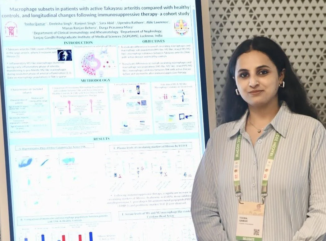
Tooba Qamar is a PhD scholar (DST-INSPIRE Fellow) in the Department of Clinical Immunology and Rheumatology at Sanjay Gandhi Postgraduate Institute of Medical Sciences (SGPGIMS), Lucknow, Uttar Pradesh. Her research focuses on vascular rheumatology, particularly Takayasu arteritis, with an emphasis on macrophage polarization, vascular inflammation, and fibrosis.
Linkedin: www.linkedin.com/in/tooba-qamar-7286341ab
Lab PI name: Dr Durga Prasanna Misra (Additional Professor of Clinical Immunology and Rheumatology, SGPGIMS, Lucknow)
University: Department of Clinical Immunology & Rheumatology, Sanjay Gandhi Postgraduate Institute of Medical Sciences (SGPGIMS), Lucknow-226014, U.P., India
What was the core problem you aimed to solve with this research? Takayasu arteritis (TAK) is a rare, stenosing large vessel vasculitis characterized by arterial wall fibrosis. Longitudinal changes in circulating monocyte/macrophage populations, which drive both inflammation and fibrosis in TAK, have not been studied. Therefore, the core problem our research addressed was the limited understanding of how inflammation and blood vessel scarring are linked in Takayasu arteritis (TAK). While immunosuppressive therapy is used to control inflammation, its relationship with the immune cells and biological processes associated with arterial fibrosis remains unclear. By comparing patients with active TAK with healthy controls and tracking changes following immunosuppressive therapy, we identified changes in M1-like and M2-like monocyte/macrophage populations, their associated cytokines, and circulating markers of fibrosis. This highlights an important gap in current disease management: controlling inflammation may not be sufficient to address the processes associated with arterial fibrosis. Our research provides a foundation for further investigating whether combining immunosuppressive therapy with anti-fibrotic treatments could offer a more comprehensive approach to protecting blood vessels in patients with TAK.
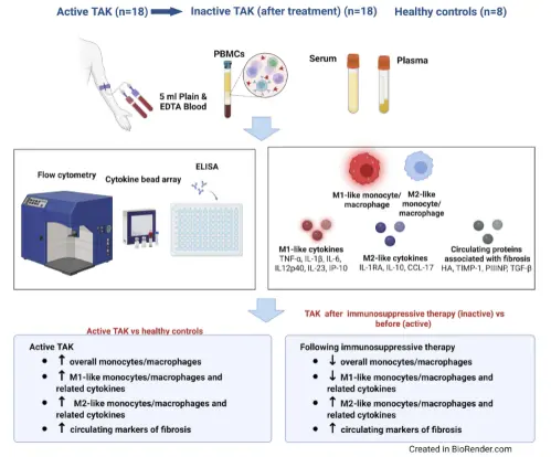
How did you go about solving this problem? To address this problem, we longitudinally evaluated circulating monocyte/macrophage populations, their associated cytokine profiles, and circulating markers of arterial wall fibrosis in patients with active Takayasu arteritis (TAK), with healthy controls serving as the reference group. Peripheral blood mononuclear cells were analyzed by flow cytometry to characterize the overall CD14+CD66b− monocyte population and identify M1-like and M2-like subsets. Serum M1-associated cytokines (TNF-α, IL-1β, IL-6, IP-10, IL-12p40, and IL-23) and M2-associated cytokines (IL-1RA, IL-10, and CCL17) were quantified using cytokine bead array or ELISA. In parallel, circulating markers associated with arterial wall fibrosis, including hyaluronic acid (HA), procollagen type III N-terminal propeptide (PIIINP), tissue inhibitor of metalloproteinase-1 (TIMP-1), and transforming growth factor-beta (TGF-β), were measured using ELISA. This integrated approach enabled us to investigate the relationship between monocyte/macrophage polarization, inflammatory mediators, and fibrotic processes in TAK over time.
‘’Our findings underscore the need to investigate combined immunosuppressive and anti-fibrotic strategies to address inflammation and vascular fibrosis in Takayasu arteritis’’ – Dr Durga Prasanna Misra
How would you explain your research outcomes (Key findings) to the non-scientific community? Our research focused on Takayasu arteritis (TAK), a rare cardiovascular autoimmune disorder in which the immune system attacks large blood vessels, causing inflammation, damage, and scarring. Compared with healthy controls, patients with active TAK had higher levels of immune cells associated with inflammation (M1-like macrophages) and tissue repair (M2-like macrophages). We also found increased levels of inflammatory cytokines, including TNF-α, IL-6, and IL-23, and proteins associated with blood vessel scarring, such as hyaluronic acid (HA) and PIIINP. Interestingly, following immunosuppressive therapy, M1-like macrophages and several inflammatory cytokines decreased, suggesting that treatment helped reduce inflammation. However, M2-like macrophages, cytokines associated with immune regulation and tissue repair (including IL-10 and CCL17), and several markers linked to blood vessel scarring (HA, TIMP-1, PIIINP, and TGF-β) increased. These findings suggest that although immunosuppressive therapy helps control inflammation, processes associated with blood vessel scarring may persist or become more prominent. This highlights the need to explore treatments that target both inflammation and fibrosis. To our knowledge, this is the first study to investigate changes in M1-like and M2-like monocyte/macrophage populations, their associated cytokines, and circulating markers of fibrosis in patients with TAK over time. One limitation was that we could not directly examine immune cells within blood vessel walls because obtaining arterial tissue usually requires major surgery. The study also involved a relatively small number of patients, and blood protein measurements can naturally vary. Ultimately, our findings support further investigation into combining immunosuppressive therapy with potential anti-fibrotic treatments to improve long-term outcomes for people living with TAK.
What are the potential implications of your findings for the field and society? To conclude, this study reports for the first time an elevated overall monocyte/macrophage population and M1-like or M2-like sub-populations, and their related cytokines and circulating fibrotic proteins in patients with TAK than healthy controls. Following the initiation of immunosuppressive therapy, the circulating monocyte/macrophage populations and cytokines showed a transition to a dominant M2-like phenotype and a profibrotic milieu indicated by elevated levels of fibrotic proteins which could indicate a reparative phase that if goes unchecked, might result in permanent arterial scarring. Targeting M2-like monocytes/macrophages and fibrosis in patients with TAK after they attain inactive disease merits exploration in pre-clinical models and eventually in clinical trials.
What was the exciting moment during your research?
The most exciting moment during our research was discovering that, although immunosuppressive therapy reduced inflammation in patients with Takayasu arteritis (TAK), several markers associated with blood vessel scarring increased. We observed a decrease in inflammation-associated immune cells (M1-like macrophages) and their related cytokines, alongside an increase in repair-associated immune cells (M2-like macrophages) and markers of fibrosis. This unexpected finding revealed that the biological processes associated with inflammation and scarring may follow different patterns during treatment. Seeing these changes emerge through the longitudinal assessment of patients, compared with healthy controls, was particularly exciting because it opened a new avenue for understanding why blood vessel damage may remain a concern even when inflammation improves. It strengthened our motivation to explore whether combining immunosuppressive therapy with anti-fibrotic approaches could offer better protection against vascular damage in TAK.
Paper reference/citation (with link): Qamar T, Singh D, Chauhan RS, Abid S, Rathore U, Lawrence A, Behera MR, Jain N, Ora M, Misra DP. Monocyte/Macrophage Subsets in the Peripheral Blood, Their Related Cytokines, and Circulating Markers of Fibrosis in Active Takayasu Arteritis and Longitudinal Changes Following Immunosuppressive Therapy. J Inflamm Res. 2026;19:602674, https://doi.org/10.2147/JIR.S602674.