



Extracellular Vesicle-associated Cofilin-1: A Promising biomarker for Parkinson’s Disease
Research Summary: This study investigates extracellular vesicle-associated Cofilin-1 (CFL1) as a minimally invasive candidate for the diagnosis of Parkinson’s disease (PD). EV-CFL1 was significantly elevated in PD patients and showed promising diagnostic potential to differentiate PD from progressive supranuclear palsy (PSP). Evidence from an in-vivo PD mouse model and human PD brain tissue analysis further supported the association of CFL1 with PD pathology.
Researcher Spotlight
Ms. Deepthy Chandran is a PhD research student working under the guidance of Dr. Srinivas G. in the Department of Biochemistry, SCTIMST. Her PhD work is on identification and characterization of extracellular vesicle cargoes in Parkinson’s disease.
Linkedin: https://www.linkedin.com/in/deepthy-chandran-a-3145a9a4/
Twitter: @deepthy_ca
Instagram: deepthy6319
Lab PI name: Dr. Madhusoodanan U.K.
University/Institute: Sree Chitra Tirunal Institute for Medical Sciences and Technology (SCTIMST), Trivandrum, Kerala
What was the core problem you aimed to solve with this research?
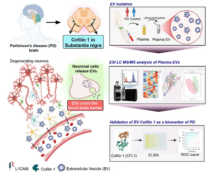
Despite extensive research on biomarkers for PD diagnosis, there remains a lack of reliable and less-invasive biomarkers for its early diagnosis and differentiation from atypical Parkinsonian disorders such as PSP. Since clinical diagnosis of PD often occurs after substantial neuronal loss, there is a need for biomarkers that can provide information about pathological changes in the brain at an earlier stage. We focused on extracellular vesicles (EVs) because they are released by cells and carry proteins and other molecular cargo that reflect the biological state of their cells of origin. Importantly, EVs are present in accessible biofluids such as plasma and can cross the blood-brain barrier, a minimally invasive source of molecular signatures that could reflect changes occurring in the brain. We investigated whether EVs in plasma could serve as a source of disease-associated biomarkers. Specifically, this study focused on EV-CFL1 to evaluate its potential for distinguishing PD from healthy controls and PSP, and to explore its relevance as a biomarker.
How did you go about solving this problem?
We approached this problem through a combination of proteomic discovery, clinical validation using ELISA assays, and other complementary experiments. First, isolated EVs from plasma using ultracentrifugation and characterized them based on their size, morphology, and EV-specific marker proteins. Next, untargeted proteomic analysis was performed to identify proteins associated with PD, and the identified proteins were compared with established EV databases, including Vesiclepedia and ExoCarta. This analysis identified Cofilin-1 (CFL1) as a promising candidate. The proteomic findings were validated using ELISA in an independent cohort, comparing EV-CFL1 levels among patients with PD, PSP, and healthy controls. The diagnostic performance of the tests was evaluated using ROC and precision-recall curve analyses and examined its association with age, disease severity, and α-Synuclein levels. Finally, to investigate the biological origin and relevance of EV-CFL1, L1CAM-based immunoprecipitation was performed to enrich brain-derived EVs, which showed that the CFL1 is enriched in LICAM positive EVS suggesting that they are originating from the brain. We further examined CFL1 expression in a mouse PD model and human PD brain tissue using immunofluorescence and immunohistochemistry, respectively. Overall, this integrated approach allowed us to investigate EV-CFL1 and its relevance to PD pathology.
“Our study identified EV-CFL1 as a potential biomarker for early diagnosis of Parkinson’s disease (PD) and, importantly, for distinguishing PD from progressive supranuclear palsy (PSP), addressing a major challenge in early clinical diagnosis and treatment. The increased presence of CFL1 in the PD brain highlights its potential role in disease pathogenesis, opening new avenues to develop CFL1-targeted disease-modifying therapies”. -Dr. Madhusoodanan U.K.
How would you explain your research outcomes (Key findings) to the non-scientific community?
PD can be difficult to identify in its early stages, when changes are already taking place in the brain. This led us to ask a simple question: Can a blood sample give us clues about what is happening inside the brain? We focused on tiny particles in the blood called extracellular vesicles (EVs). These vesicles act like small packages released by cells. They carry proteins and other molecular information from their cells of origin, providing insights into changes occurring in the brain. We found that one protein, Cofilin-1 (CFL1), was present at significantly higher levels in the EVs of PD patients compared with healthy individuals. Interestingly, CFL1 levels were also higher in PD than in PSP, a condition that can have symptoms similar to PD. This finding suggests that CFL1 may have potential to help distinguish between these two conditions. We then looked for evidence that the CFL1 we detected in the blood was related to changes occurring in the brain. Our findings in brain tissue from PD patients and in a PD mouse model supported this possibility.
Overall, our study suggests that CFL1 carried by blood EVs could provide a glimpse into disease-related changes in the brain through a simple blood sample.
What are the potential implications of your findings for the field and society?
From a scientific perspective, this work highlights the potential of EVs as a window into pathological changes occurring in the brain. Cofilin-1 (CFL1) is an actin-binding protein that regulates the actin cytoskeleton, which is important for maintaining the neuronal structure and function. Altered cofilin regulation may contribute to neuronal dysfunction in PD, making the identification of CFL1 in plasma EVs relevant for understanding PD-associated molecular changes.
From a clinical perspective, EV-CFL1 could potentially contribute to a minimally invasive blood-based approach for PD diagnosis and distinguishing PD from PSP. This could ultimately complement current clinical and neurological assessments.
However, the study emphasizes that EV-CFL1 is currently a candidate biomarker for PD rather than an established diagnostic test and larger, longitudinal studies are needed to determine its clinical as well as diagnostic relevance.
What was the exciting moment during your research?
A key finding of this study was the independent validation of the proteomic discovery of EV-CFL1, which demonstrated significantly elevated EV-CFL1 levels in PD patients compared with healthy controls. This validation provided further evidence for the reproducibility of the proteomic findings and the potential of EV-CFL1 as a candidate biomarker for PD.
It became even more interesting when we found that CFL1 could differentiate PD from PSP, suggesting that it might have disease-specific diagnostic relevance. Finally, demonstrating CFL1 in plasma EVs from PD patients, and observing increased CFL1 expression in the substantia nigra of PD models and human PD brain tissue, provided an important biological link between our blood-based finding and the pathology occurring in the brain.
The most rewarding aspect in our study was seeing the research progress from an unbiased proteomic discovery to independent clinical validation and biological evidence supporting its relevance to PD.
Paper reference: Cofilin 1 is an Extracellular Vesicle-Associated Plasma Biomarker of Parkinson’s Disease in Humans. Deepthy Chandran, Unnati Agrawal, Deepa Damayanthi, Divya T. Nair, Geetha Mandagini, Sruthi S. Nair, Deepti Narasimhaiah, Francis Boniface Fernandez, Poonam Thakur, Cibin T. Raghavan, Syam Krishnan, Srinivas Gopala, Madhusoodanan Urulangodi. Annals of Neurology (2026); 00:1-18. https://doi.org/10.1002/ana.78348