



IP3R2: The Ca2+ checkpoint for melanosome degradation
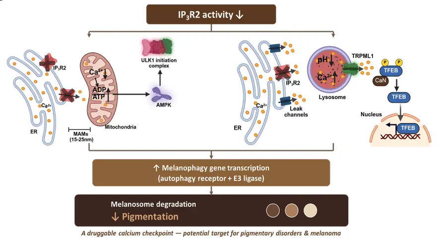
Research Summary: IP3R2 loss reduces mitochondrial Ca²⁺ uptake and elevates lysosomal Ca²⁺ via TMEM165. This activates TRPML1–TFEB–RCHY1/OPTN signaling, promoting melanosome degradation and reducing pigmentation. Hence, IP3R2 restricts melanosome degradation.
Researcher Spotlight
Suman Saurav recently completed his PhD under supervision of Dr. Rajender Motiani at RCB, Faridabad. His work revealed how IP₃R2 regulates inter-organelle calcium signaling to control melanosome degradation.
Linkedin: www.linkedin.com/in/suman-saurav-phd-77840a118
Twitter: https://x.com/sumansaura86737
Instagram: https://www.instagram.com/suman_saurav_1/
Lab PI name: Dr. Rajender Motiani, Regional Centre for Biotechnology, Faridabad, India
Lab social media: https://www.linkedin.com/company/calciomicsgroup-rcb/posts/?feedView=all
What was the core problem you aimed to solve with this research?
Skin pigmentation depends on a balance between melanosome biogenesis and degradation. Melanosome biogenesis is well characterized. However, the mechanisms governing melanosome degradation (melanophagy) remain poorly understood.
Calcium (Ca²⁺) is an important regulator of pigmentation. However, how calcium signals exchanged between different cellular organelles influence melanophagy was unknown. We therefore set out to identify the calcium channel that controls melanophagy. We also investigated, how Ca²⁺ transfer between the endoplasmic reticulum, mitochondria, and lysosomes regulates the degradation of melanosomes. A further challenge was that there was no effective live-cell method to directly visualize and monitor melanophagy in real time.
We developed tools to dynamically track melanophagy in living cells. Our research addressed these gaps by identifying IP3R2 as an important regulator of inter-organelle calcium signaling and melanophagy.
How did you go about solving this problem?
Identification- Two independent microarrays (mouse melanocytes and primary human melanocytes) analysis identified IP3Rs as differentially regulated genes. IP3R2 emerged as the only IP3R isoform consistently associated with pigmentation. Isoform-specific knockdown confirmed that only IP3R2 depletion reduced melanin, across multiple cell models. Wild-type IP3R2 rescued this effect, whereas a pore-dead mutant did not, demonstrating that Ca²⁺ conductance is essential. In zebrafish, morpholino knockdown reduces melanin and IP3R2 mRNA restores it.
Melanophagy readout- We built two ratiometric live-cell probes, mCherry-Tyrosinase-EGFP and Tyrosinase-mKeimaN1. We validated them with rapamycin, BafA1, retagliptin and HMB45 co-localization, and cross-checked them with LC3-HMB45 co-localization and TEM counts of stage III/IV melanosomes.
Lysosomal dissection- IP3R2 knockdown lowered lysosomal pH and raised lysosomal Ca²⁺ (OBDx, BafA1, GPN), even after thapsigargin pre-depletion. It also increased ER Ca²⁺ leak and TMEM165 expression, and PLA and TEM showed closer ER–lysosome apposition. TMEM165 gain and loss, TRPML1 wild-type versus pore-dead rescue, and TFEB translocation with RCHY1/OPTN co-silencing established the TMEM165–TRPML1–TFEB–RCHY1/OPTN axis.
Mitochondrial arm- CEPIA2mt, ADP/ATP, p-AMPK/p-ULK1 and MCU overexpression showed that reduced mitochondrial Ca²⁺ uptake feeds melanophagy in parallel.
“This work reveals how calcium signaling fine-tunes pigmentation, positioning IP3R2 as a promising node for treating pigmentary disorders.” – Dr. Rajender Motiani
How would you explain your research outcomes (Key findings) to the non-scientific community?
Skin pigmentation is determined by the balance between the melanosome biogenesis and their clearance via melanophagy. Inside the cell, the ER acts as a calcium warehouse, and IP3R2 is a gate on that warehouse. Through this gate, calcium reaches the mitochondria, the cell’s power plants, and keeps them running. When IP3R2 is blocked, the power plants receive less calcium, ATP levels decrease relative to ADP, and the cell experiences an energy shortage. The energy sensor AMPK then activates ULK1, which starts the cleanup crew.
At the same time, the lysosomes, the recycling centers, move closer to the warehouse and take up extra calcium through a transporter called TMEM165. They also become more acidic, which switches on the calcium channel TRPML1. This releases calcium that pushes TFEB, the master switch of the recycling programme, into the nucleus. TFEB turns ‘’ON’’ two key regulators- RCHY1 and OPTN. RCHY1 tags pigment packets for disposal, and OPTN carries them to the lysosome.
So IP3R2 acts as a brake on melanosome degradation. When IP3R2 is blocked, the cell recycles its own melanosome and becomes lighter. We saw the same phenomenon in cultured cells and zebrafish.
What are the potential implications of your findings for the field and society?
For the field: This study establishes melanophagy as a Ca²⁺-regulated process. It identifies IP3R2 as a key upstream regulator. IP3R2 acts as a gatekeeper that feeds two parallel arms, and both converge on melanosome degradation:
ER–mitochondria arm: reduced mitochondrial Ca²⁺ uptake raises the ADP/ATP ratio and activates AMPK–ULK1, independent of mTOR.
ER–lysosome arm: Closer ER–lysosome apposition promotes TMEM165-mediated lysosomal Ca²⁺ loading. This activates TRPML1. TRPML1 activation drives TFEB into the nucleus, inducing RCHY1 and OPTN.
The study also provides live-cell tools to monitor melanophagy and identifies several potential regulatory nodes for future investigation.
For society: Pigmentation is our main defense against UV and its dysregulation leads to pigmentary disorders such as melasma and vitiligo. Enhancing melanosome degradation alleviates hyperpigmentation, whereas inhibiting it improves hypopigmentation. IP3R2 expression is higher in African skin than in White skin, and lightly pigmented human melanocytes have more acidic, Ca²⁺-rich lysosomes. The IP3R2–Ca²⁺ pathway provides potential targets for future therapeutic research, although these findings require validation in human disease models before any clinical application.
What was the exciting moment during your research?
The surprise came from the lysosomes. We initially expected IP3R2 loss to reduce lysosomal Ca²⁺, but multiple independent approaches, including OBDx, BafA1, and GPN consistently showed the opposite (lysosomal Ca²⁺ increased). This unexpected result became the critical clue that led us to uncover a pathway involving ER Ca²⁺ leakage, TMEM165, TRPML1, and TFEB, ultimately linking inter-organelle Ca²⁺ communication to melanophagy.
Paper reference: Saurav S, Jadon A, Motiani RK. (2026). IP₃R2-mediated inter-organelle calcium signaling suppresses melanosome degradation. PLoS Biol. 2026 Aug 25;24(8):e3003971. https://doi.org/10.1371/journal.pbio.3003971