



LncRNA Lockd Fuels Cardiomyocyte Proliferation to Protect the Stressed Heart
Research Summary: Neonatal hearts during the regenerative window resist pathological stress. LncRNA Lockd overexpression promotes regeneration beyond the regenerative window by stabilizing ENO1 to activate β-catenin/Myc signaling and drive cardiomyocyte proliferation.
Researcher Spotlight
First authors: Dr. Shakti Prakash and Samprikta Kundu (equal contribution)
Dr. Shakti Prakash is a cardiovascular molecular biologist who completed a Ph.D. at CSIR-CDRI, Lucknow, studying long non-coding RNAs, AAV-based gene therapy and cardiac regeneration.
LinkedIn: https://www.linkedin.com/in/shakti-prakash-ph-d-463464114/.
Google Scholar: https://scholar.google.com/citations?user=V7xT73cAAAAJ&hl=en.
Lab PI name: Dr. Shashi Kumar Gupta, Principal Scientist, Pharmacology Division
Institute: CSIR-Central Drug Research Institute (CSIR-CDRI), Lucknow.
Lab website https://www.linkedin.com/in/shashi-kumar-gupta-41105187/
What was the core problem you aimed to solve with this research?
Heart disease is the leading cause of death worldwide, largely because the adult human heart cannot replace the heart muscle cells (cardiomyocytes) it loses after injury or long-term stress. Instead, the heart thickens (hypertrophy), stiffens and eventually fails. Interestingly, newborn mouse hearts can regenerate during the first week of life, but this ability is lost soon afterwards. This regenerative window has been well studied using injury models such as apical resection (surgical removal of the heart tip) and myocardial infarction (heart attack), and using pressure overload by aortic banding. However, the response of the newborn heart to isoproterenol, a widely used drug that induces pathological cardiac hypertrophy, had not been studied during the neonatal stage. We therefore decided to treat newborn mice with isoproterenol, both during and after the regenerative window, to answer two questions: does the regenerative window also protect the heart from isoproterenol-induced hypertrophy, and if so, which molecular switch keeps heart muscle cells dividing during this period? Finding that switch could point to ways of reawakening regeneration in the adult heart.
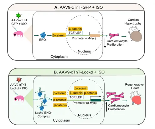
Figure Caption: How Lockd keeps heart muscle cells dividing. (A) In control, where heart muscle cells have stopped dividing, hearts exposed to the stress-inducing drug isoproterenol become enlarged (hypertrophy). (B) When Lockd is delivered to heart muscle cells using a heart-specific AAV9 vector, Lockd binds to and protects the protein ENO1. This increases β-catenin, which moves into the nucleus and switches on the growth gene c-Myc. As a result, heart muscle cells keep dividing, and the heart stays healthy under stress.
How did you go about solving this problem?
We treated newborn mice with isoproterenol, a drug that drives cardiac hypertrophy, either during the regenerative and non-regenerative phase. We compared heart size, cardiomyocyte size, heart function by echocardiography, and the proliferation markers Ki67, Aurora B and pH3 between the two groups. We then performed RNA sequencing of regenerative and non-regenerative hearts to look for molecules that could explain the difference, and selected the long non-coding RNA Lockd for further study. To test its function, we used a heart-specific AAV9-cTnT gene-delivery vector to keep Lockd switched on in hearts beyond the regenerative window. To uncover the mechanism, we pulled Lockd out of heart cells and identified its protein partners by mass spectrometry, then tested its binding to ENO1 by RNA immunoprecipitation, recombinant-protein pull-down and RNA-EMSA. Finally, we used protein-stability assays, shRNA knockdown and confocal imaging to study the ENO1/β-catenin/Myc pathway.
“This study shows how a non-coding RNA keeps young heart cells dividing, opening new avenues for cardiac regeneration.” — Dr. Shashi Kumar Gupta
How would you explain your research outcomes (Key findings) to the non-scientific community?
Our hearts are built from muscle cells that, in adults, cannot be replaced once lost. Newborn hearts are different: for a short time after birth, they have an active “repair team” that can make new heart muscle cells. We found that when newborn mouse hearts were stressed with isoproterenol during this window, they simply made more cells and kept working normally. Just a few days later, once the repair team had gone quiet, the same stress made the heart cells swell, the heart enlarged, and it began to fail- a condition called cardiac hypertrophy and heart failure. We discovered that a molecule called ‘Lockd’ acts as a leader of this repair team. When we kept Lockd active in older baby mouse hearts, the heart cells continued dividing, and the heart remained healthy under hypertrophic stress. Lockd does this by protecting a protein called ENO1, which switches on growth signals (β-catenin and Myc) that tell heart cells to divide.
What are the potential implications of your findings for the field and society?
For the field, our study adds Lockd to a small but growing list of long non-coding RNAs that control heart muscle cell proliferation. It also reveals a non-enzymatic role for ENO1, a protein best known as a sugar-metabolism enzyme, as an RNA-binding partner that controls β-catenin signalling. In addition, it shows that the regenerative window of the newborn heart protects it against drug-induced hypertrophy, not only against physical injury. For society, the long-term hope is that therapies which reactivate pathways like Lockd/ENO1/β-catenin/Myc could help damaged adult hearts repair themselves rather than fail. Because we delivered Lockd with a heart-targeted AAV9 vector, the work also connects to the growing field of cardiac gene therapy. However, these experiments were done in newborn mice and cultured cells. Testing whether this pathway can be activated in adult hearts, for example after a heart attack, and whether a similar mechanism operates in humans are the essential next steps before any clinical application.
What was the exciting moment during your research?
Instead of studying aging or failing hearts, as most research does, we focused on the relatively unexplored transition stage, when the newborn heart loses its ability to regenerate. Our reasoning was simple: if we could find what switches regeneration off, we might find real targets to switch it back on. The most exciting moment came when we harvested the hearts from the Lockd experiment. The isoproterenol-treated control hearts were clearly enlarged, while the Lockd hearts looked almost like healthy ones, and the echocardiography and staining results confirmed that the heart cells were dividing and the hearts were pumping normally. Seeing a single non-coding RNA make such a visible difference to a whole organ told us our focus on the transition stage was the right one.
Paper reference: Prakash S, Kundu S, Chaudhary AD, Chaudhary S, Kumar C, Kumari S, Singh S, Yadav B, Kumar A, Prajapati VM, Jagavelu K, Kumar S, Gupta SK. lncRNA Lockd promotes cardiomyocyte proliferation via Eno1/beta-catenin/Myc axis. Molecular Therapy: Nucleic Acids. 2026;37:103132. https://doi.org/10.1016/j.omtn.2026.103132.