



Room to Grow: How Physical Confinement Paces Yeast Cell Division
Research Summary: Growing budding yeast inside soft, transparent 3D hydrogels, we found that physical confinement alone slows cell division by restricting bud growth during G2, with no detectable mechanosensory or stress signatures.
Researcher Spotlight
First authors. Sreepadmanabh and Mridul Gautam (joint first authors)
M Sreepadmanabh (Sree) is a PhD student in the Bhattacharjee lab at NCBS, where his doctoral research explores how complex 3D environments regulate living systems across scales.
Linkedin | Twitter | Instagram
Mridul Gautam is a Master’s student at TH Mannheim. During his stint in the Bhattacharjee lab, he investigated how 3D physical confinement shapes cellular growth and division.
Lab PI name: Dr. Tapomoy Bhattacharjee
University: National Centre for Biological Sciences, Tata Institute of Fundamental Research (NCBS-TIFR), Bengaluru, India
Lab social media: https://www.esoftbio.com/
What was the core problem you aimed to solve with this research?
Almost everything we know about how a cell divides comes from cells grown in conditions that would look rather strange to the cells themselves: swimming freely in a flask of nutrient broth or sitting on the flat surface of an agar plate. Out in the real world, life is rarely so roomy. Microbes live packed into soil, wedged within mucus, embedded inside tissues and crowded into biofilms. These habitats are three-dimensional, porous and squishy – soft materials that hold cells in place and push back on them as they grow.
Budding yeast (Saccharomyces cerevisiae) has been one of biology’s great workhorses for understanding the cell cycle – the tightly choreographed sequence of growth, DNA replication, and division that every cell goes through. Decades of elegant genetics have revealed the molecular machinery that drives this cycle: cyclins, kinases, checkpoints and mechanisms that let a cell sense its own size. But that left us with a question we found hard to ignore: can a cell’s physical surroundings, on their own, change how fast it divides?
Answering this turned out to be trickier than it sounds. Earlier studies had grown yeast under confinement in microfluidic chambers – and in some of these set-ups, densely packed, growing cells build up large compressive stresses and show abnormal shapes or arrested growth. However, none of these platforms really capture the porous, soft, and viscoelastic character of natural 3D environments. Just as importantly, many of the forces that cells encounter in nature are often gentle – frequently below the levels expected to trigger classical mechanosensing pathways. So, we set out to ask whether in a more realistic, soft 3D environment, could purely physical confinement regulate something as molecularly orchestrated as the cell cycle?
How did you go about solving this problem?
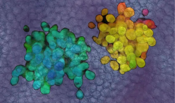
This is where soft matter physics meets cell biology. Our lab builds engineered 3D environments out of granular hydrogels. Imagine a jar packed tightly with microscopic, highly swollen, jelly-like beads. (Fun fact: the polymer we use, Carbopol, belongs to the same family used to thicken hand sanitisers and hair gels.) Because the beads are jammed together, the packing behaves like a soft solid – it holds its shape and keeps cells firmly in place. But push on it hard enough, beyond a point called its yield stress, and the beads locally rearrange and flow. So, a growing yeast colony can push its way outwards, while being continuously resisted by the surrounding matrix. Best of all, the material is transparent, which means we can watch individual cells bud and divide in real time under the microscope, deep inside a 3D environment.
We prepared these matrices directly in yeast growth medium and tuned how tightly the beads were packed, creating environments with different degrees of confinement. Using rheology – the science of how soft materials deform and flow – we characterized them carefully. Our ‘low confinement’ matrix has a yield stress of about 3 Pa and an elastic modulus of about 30 Pa; our ‘high confinement’ matrix has a yield stress of about 60 Pa and a modulus of about 330 Pa. To put that in perspective, these materials are hundreds of times softer than the pressure that inflates a yeast cell from within, and more than a thousand times gentler than the pressures applied in earlier experiments. We even made an ‘ultra-low confinement’ matrix, with a yield stress of only about 0.24 Pa – and strikingly, even that was enough to slow down yeast growth.
We then measured how quickly whole populations grew, tracked colony sizes over several days and, most importantly, filmed individual mother cells to time exactly how long each took between producing successive buds. To pinpoint where in the cell cycle any delay was happening, we used yeast strains carrying fluorescently tagged ‘reporter’ proteins that mark specific stages – the phosphatase Cdc14, the septin Cdc10 and the spindle pole body component Spc42. Combined with careful measurements of how fast buds grew, these let us dissect G1, S phase, G2 and anaphase separately, cell by cell.
Of course, a slower cell cycle can have many possible explanations, so we invested a great deal of effort in ruling them out. We raised the osmotic pressure of liquid media up to 100-fold above the elastic modulus of our high-confinement matrix, controlled for pH, examined cell shape and mitochondrial morphology, and profiled both the transcriptome (using RNA-seq) and the proteome. We also tested yeast strains lacking each of eight well-studied mechanosensing and mechanotransduction genes – HOG1, WSC1, WSC2, WSC3, MID2, MYO1, TPM1 and TPM2 – to see whether any of them was responsible for biochemically altering the cellular behavior under confinement. All of this was only possible due to a dedicated joint effort, working alongside our colleague Nikita Bagade at Dr. Tapomoy Bhattacharjee’s lab, and in collaboration with Prof. Sunil Laxman’s group at inStem, Bengaluru.
“Our findings invite a re-examination of how we traditionally study living systems. Most laboratory culture conditions strip away the mechanical complexity of real habitats. By restoring that complexity in a controllable way, our work has shown that purely physical variables can act as primary regulators of fundamental biological processes.” – Dr. Tapomoy Bhattacharjee
How would you explain your research outcomes (Key findings) to the non-scientific community?
Picture a yeast cell as a tiny balloon that reproduces by inflating a smaller balloon on its side – the daughter ‘bud’. The bud grows until it is big enough, and then separates as a brand-new daughter cell. In a flask of broth, nothing gets in the way, and under ideal lab conditions a yeast cell can complete this cycle in roughly an hour and a half.
Now imagine trying to inflate that balloon while it is buried in a jar of very soft jelly. The jelly doesn’t crush the balloon, and it doesn’t damage it – but every bit of growth now has to nudge the jelly out of the way. That is essentially what we saw. In our soft 3D matrices, yeast cells took noticeably longer between producing successive buds because they kept growing slower, and the more confining the matrix, the longer the wait and the slower the growth.
What surprised us most was everything that did not happen. The cells looked perfectly healthy. Their shape and size were normal, their mitochondria – the cell’s powerhouses – looked unchanged, and populations eventually reached the same final sizes as in liquid culture; they just took longer to get there. When we compared cells under high and low confinement, they were remarkably similar at the level of their genes and proteins, with no sign of stress responses or mechanosensing programmes being switched on. Yeast strains, each missing one of eight different ‘force-sensing’ genes behaved just like normal yeast – they were slowed down by confinement too. And most strikingly, when we pulled cells out of the jelly and returned them to liquid broth, they immediately went back to dividing at their normal pace. The cells weren’t sick, stressed or permanently changed. They were simply short of room.
So where exactly was the hold-up? By timing each stage of the cell cycle separately, we found that most stages – the initial growth phase before DNA copying (G1), copying the DNA (S phase) and pulling the chromosomes apart (anaphase) – took the same time regardless of confinement. The delay was concentrated in one stage, G2, when the bud does much of its growing. In confined cells, buds inflated more slowly, yet they still grew to their usual size before division – so slower inflation translated directly into a longer cell cycle.
In short, the physical environment by itself – without any detectable molecular signal – can set the pace of cell division.
What are the potential implications of your findings for the field and society?
For cell biology, textbook models describe how cells decide when to divide using internal ‘sizers’ and ‘timers’. Our results show that, operating alongside these internal controls, the surroundings can directly govern how quickly a cell completes its cycle – through simple physics, apparently without the cell needing to actively sense anything. Because the stresses involved here are tiny, this kind of regulation could be at work all the time in natural habitats, entirely invisible to experiments done in flasks and on plates.
There is also a practical insight for how we do experiments. Division rates, growth phenotypes and responses to treatments are routinely measured in liquid cultures. If a cell’s behaviour in its real habitat – soil, mucus, the gut or a host tissue – depends on its physical surroundings, then some of what we measure in the lab may not translate directly. Tunable, transparent 3D platforms like ours offer a way to bridge that gap without giving up precise, quantitative imaging and analyses.
Looking further ahead, many organisms that matter for human health and the environment proliferate in confined spaces: fungi and bacteria in soil, microbes living in the mucus layers of our lungs and gut, pathogens invading host tissues, and even cancer cells growing within dense tissue. Whether the same physical principle operates in these systems is an open and exciting question that we have not yet tested – but if it does, the physical environment could become a new lever for understanding, and perhaps one day steering, how fast cells proliferate. Our platform also offers a rare experimental tool: a way to slow down the cell cycle in a tunable manner, without drugs, genetic modifications or nutrient starvation. That opens up fundamental questions of its own – for instance, how slower division might influence ageing in yeast, which is itself a classic model for ageing research.
We should also be clear about what our study does not show. The environments we used are very soft; in stiffer surroundings, where stresses can build up locally around a growing bud, cells may well engage their mechanosensing and stress pathways. And although we found no signatures at the level of gene expression or protein abundance, we cannot rule out subtler changes – for example, in where proteins sit inside the cell or how they are chemically modified. Untangling these possibilities is a natural and exciting next step.
What was the exciting moment during your research?
For us, there were really two moments that stood out. The first was a moment of ‘wait – nothing changed?’ When the transcriptomics data came back, we half-expected to find a dramatic stress or mechanosensing signature that would explain why confined cells were dividing more slowly. Instead, cells under low and high confinement were almost indistinguishable. At first that felt like a dead end, but it quickly became the most exciting part of the story, because that ‘null’ result was telling us something deeper: the slowdown might not be a molecular response at all, but a direct physical consequence of the environment. The proteomics and the eight mechanosensing mutants told exactly the same story.
The second was when we took cells out of the high-confinement matrix, put them back into liquid broth, and watched them snap straight back to their normal budding rhythm. That was when it truly clicked: the cells hadn’t been damaged or reprogrammed – the slowdown was purely their surroundings. Seeing a process as molecularly sophisticated as the cell cycle being paced by something as simple as a soft jelly-like matrix and being able to watch it unfold cell by cell in real time, was genuinely thrilling.
Paper reference: M Sreepadmanabh*,#, Mridul Gautam#, Nikita Bagade, Sunil Laxman, and Tapomoy Bhattacharjee* – 3D confinement physically regulates cell cycle progression in budding yeast. Nature Communications (2026). https://doi.org/10.1038/s41467-026-78414-y. # These authors contributed equally; * corresponding authors