



Decoding tumor-innate immune cell crosstalk in cancer cachexia
Research Summary: Tumors hijack innate immune cells and hematopoiesis, driving systemic insulin resistance and tissue wasting through ImpL2-mediated disruption of the hematopoietic niche in cancer cachexia.
Researcher Spotlight
Ujjayita Chowdhury is a final year PhD student at Tata Memorial Centre-ACTREC wrapping her head around inter-organ communication in the Drosophila model of cancer cachexia.
LinkedIn: https://www.linkedin.com/in/ujjayita-chowdhury-7673aa208
Twitter: https://x.com/UjjayitaC
Instagram: https://www.instagram.com/el_condor_pasa_15
Lab PI name: Dr. Rohan J. Khadilkar
University: Tata Memorial Centre- Advanced Centre for Treatment Research and Education in Cancer (TMC-ACTREC)
Lab social media: https://scthlab.wixstudio.com/scthlab
What was the core problem you aimed to solve with this research?
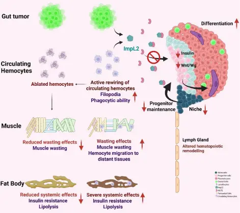
Cancer cachexia is a multi-organ syndrome that is irreversible, and it is prevalent in around 80% of cancer patients in their advanced stages. The role of blood cells or formation of blood cells i.e. hematopoiesis in cancer cachexia is fairly under characterized. The core problem we identified over the course of my doctoral journey was elucidating how epithelial tumors signal to rewire the hematopoietic system and circulating immune cells. This was particularly relevant in a condition like cancer cachexia, which is primarily how tumors grow at the expense of the energy balance in the major host organs. It was unknown whether tumors secrete molecules that regulate hematopoietic homeostasis. We set out to address whether the tumors re-educate the innate immune cells to bring about distant systemic inflammation.
How did you go about solving this problem?
We approached this problem using a Drosophila larval midgut tumor model combining genetic manipulation, cellular imaging, functional assays, and transcriptomic profiling to investigate tumor–immune interactions. We induced intestinal tumors by activating mutations in the adult midgut precursors (AMPs) of the larval midgut and examined their systemic effects in skeletal muscles, larval fat body (similar to mammalian adipose tissue), the hematopoietic organ and circulating blood cells.
We characterized lymph gland architecture, blood cell differentiation, hemocyte morphology, phagocytic activity, and inflammatory gene expression. We also identified the overall gene signature of the circulating blood cells in the larval cachectic model using bulk transcriptomics to delineate the factors that are upregulated in the circulating blood cells. We also ablated the blood cells using genetic techniques and addressed what are the effects of blood cell ablation in a cachexia scenario.
“This study from our lab is particularly exciting as it highlights the role of macrophages as perpetrators in the metabolic dysfunction that occurs during cancer cachexia.” – Dr. Rohan J. Khadilkar
How would you explain your research outcomes (Key findings) to the non-scientific community?
Our research reveals that cancer affects not just the organ where a tumour develops but also its macro-environment. It can send signals throughout the body, disrupting metabolism and weakening healthy tissues.
We found that tumors can reprogram immune cells circulating in the body, making them active contributors to cancer-associated wasting. One important molecule, ImpL2, acts like a brake on insulin signalling, disrupting the normal functioning of blood cell-producing tissues and contributing to metabolic imbalance.
Interestingly, we discovered that both tumour cells and immune cells contribute to this process. Reducing these signals helped restore several cachexia-associated changes. In simple terms, our research shows that tumours do not act alone; they have a macro-environment and there is a crosstalk between healthy host organs and the tumor.
What are the potential implications of your findings for the field and society?
Decoding inter-organ communication in a multiorgan syndrome like cachexia is the way forward. Various factors have been identified in the past decade; however, there is still no therapeutic regimen available in the clinical setting. It is extremely difficult to replicate a complex syndrome like cancer cachexia in higher mammalian models. Hence, simple model systems like Drosophila give us the advantage of sophisticated genetic perturbation wherein we can genetically manipulate certain genes in a particular host organ or tumor and observe the effects in several other organs.
Our findings highlight a previously underappreciated role of innate immune cells in driving cancer-associated wasting. Traditionally, cancer cachexia has been studied primarily in the context of muscle and fat loss. Our work expands this understanding by showing that tumours can also reprogram circulating immune cells and disrupt blood cell development, contributing to systemic metabolic dysfunction.
Ultimately, our work encourages a broader view of cancer: treating the tumour alone may not be enough; we also need to understand and address how the tumour alters the rest of the body.
What was the exciting moment during your research?
One of the most exciting aspects of this research was microscopy! There is something incredibly fascinating about visualizing what is happening inside an organism and seeing your hypothesis take shape under the microscope. After months of trouble shooting, I couldn’t observe the muscle wasting in our larval model of cachexia. However, the euphoric moment arrived on a rather mundane week day when I zoomed in more than what was usually done to visualize larval skeletal muscles and I could observe a beautiful muscle ‘thinning’ pattern which we later went on to find ways to analyze. This was an exciting exercise. The excitement led me to further do some other experiments to finally observe that these wasted skeletal muscles were infiltrated by blood cells and this was pretty much the turning point in our story to actually understand that there is indeed active recruitment of these blood cells, or what we call plasmatocytes, in both the tumor and the host organs.
Paper reference: Chowdhury, U., Panzade, G., Karnik, P.N., Birwadkar, P., Wadhwa, T. and Khadilkar, R.J. (2026), Drosophila midgut tumor-induced insulin resistance systemically remodels lymph gland hematopoiesis during cancer cachexia. FEBS J. https://doi.org/10.1111/febs.70741