



How Sequence Context Shapes Intrinsically Disordered Viral Protein Aggregation
Research Summary: We show that seven closely related intrinsically disordered henipavirus proteins can follow distinct aggregation pathways and form structurally different fibrils because sequence context around a conserved amyloidogenic core matters.
Researcher Spotlight
First Author: Harshita Sawdekar
Thanks to a PhD fellowship from the Infectiopôle Sud (Marseille, France), Harshita Sawdekar is currently a PhD student at Aix-Marseille Université, Marseille, France, under the joint direction of Sonia Longhi (Supervisor)1 and Joseph Chamieh (Co-supervisor)2, where she studies protein aggregation and fibrillation in viral IDPs. She completed her BS-MS in Chemical Sciences from IISER Mohali, India, in 2023.
Social media (link)
LinkedIn: Harshita Sawdekar
Twitter: @Harshita3110
Instagram: @ardhi_punjabi
ORCID: https://orcid.org/0009-0007-8219-2817
Lab PIs names:
- AFMB (Architecture et Fonction des Macromolécules Biologiques)
- IBMM (Institut des Biomolécules Max Mousseron)
University:
- Aix-Marseille Université, CNRS, AFMB, Marseille, France;
- Université de Montpellier, IBMM, Montpellier, France.
Lab social media:
LinkedIn: Laboratoire AFMB
What was the core problem you aimed to solve with this research?
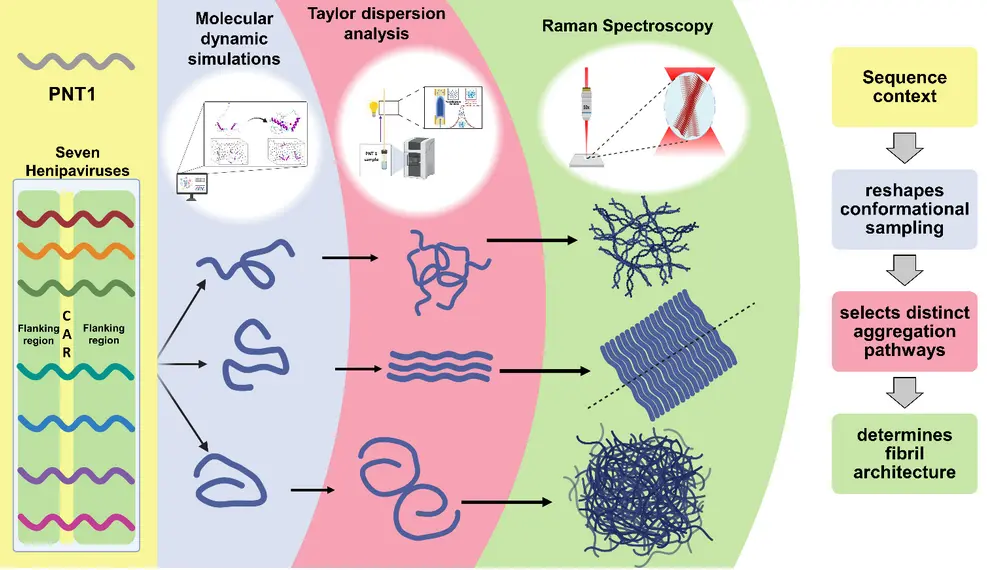
Protein aggregation in viruses remains a relatively emerging and underexplored area of research. Although intrinsically disordered proteins (IDPs) are now recognized as widespread components of viral proteomes, their ability to undergo self-assembly and form amyloid-like structures has only more recently begun to attract attention. The discovery that intrinsically disordered regions (IDRs) in viral proteins can undergo aggregation raises important questions about the molecular mechanisms that govern this process. Understanding how these proteins transition from disordered monomers to oligomeric and fibrillar assemblies, and how sequence and structural features influence these pathways, is therefore essential. In particular, elucidating the mechanisms of viral protein aggregation may ultimately help determine whether these aggregates are simply by-products of protein misfolding or whether they have functional roles in viral replication, host-pathogen interactions, and pathogenicity. In this study, we compared seven homologous PNT1 subdomains from Hendra, Nipah, Ghana, Cedar, Angavokely, Mòjiang, and Langya viruses. We combined Taylor dispersion analysis to resolve early aggregation kinetics and transient intermediates, Raman spectroscopy to characterize mature fibril structure, and all-atom molecular dynamics simulations to examine conformational flexibility and sequence-dependent structural landscapes.
How did you go about solving this problem?
A previous study from our lab identified a conserved cryptic amyloidogenic region (CAR) within the first 110 amino acids of the P, V, and W proteins across seven phylogenetically related henipaviruses. Given the conservation of this amyloidogenic core, we initially expected these proteins to exhibit broadly similar aggregation behaviors. Surprisingly, however, they followed remarkably distinct aggregation pathways: some viral proteins form transient or metastable assemblies, others follow more controlled oligomerization pathways, and some efficiently progress towards highly ordered fibrils. In addition, the architecture of the final fibrillar aggregates also diverges significantly across these proteins. These findings indicate that the conserved core is not the only determinant of protein aggregation and that its sequence context, particularly the flanking regions on either side of the CAR, is important. The key message is that an amyloidogenic core alone does not determine the fate of a protein; rather, the surrounding sequence context can profoundly influence its conformational landscape, assembly pathway, and ultimately the structure of the resulting fibrils.
“This study shows that a conserved amyloidogenic core can give rise to diverse aggregation behaviors through its surrounding sequence context.” – Dr. Sonia Longhi and Dr. Joseph Chamieh
How would you explain your research outcomes (key findings) to the non-scientific community?
Under certain conditions, proteins can interact with one another and gradually assemble into larger structures, sometimes forming long, fiber-like structures called amyloid-like fibrils. In viruses, these fibrillar assemblies may not simply be accidental clumps; they can potentially influence how the virus functions or causes disease. Understanding how and why viral proteins aggregate can therefore help us understand their role in viral biology. Our findings show that while a conserved amyloidogenic motif is sufficient to confer an inherent propensity for aggregation, it does not, on its own, dictate the aggregation pathway or the final structure of the resulting assemblies. In these viral proteins, even subtle variations in the regions surrounding the conserved amyloidogenic motif can alter the conformational landscape (i.e., all the different shapes that the protein can take) and interaction networks, leading to markedly different aggregation kinetics and fibril architectures. These results highlight how sequence variation outside a conserved amyloidogenic core can encode distinct structural and assembly behaviors, providing a broader framework for understanding the molecular diversity of viral protein aggregation.
What are the potential implications of your findings for the field and society?
For the field, the study highlights the importance of looking beyond short amyloidogenic motifs when trying to predict protein aggregation. It also provides a comparative framework for connecting sequence variation, conformational dynamics, early oligomerization, and mature fibril structure. For society, these findings may ultimately help clarify how sequence variation in viral proteins can influence molecular behavior relevant to viral biology, host-pathogen interactions, and pathogenicity.
What was the exciting moment during your research?
One particularly exciting moment in the study was when the Taylor dispersion data revealed that the seven PNT1 proteins, despite sharing a highly conserved amyloidogenic region, were taking remarkably different routes towards aggregation. We had initially expected their aggregation pathways to be broadly similar, if not identical, because of the conserved region. Instead, we found striking differences in their aggregation kinetics and intermediate states. What made this particularly interesting was that these differences were not isolated observations: they could be connected to distinct conformational landscapes revealed by molecular dynamics simulations and, ultimately, to differences in the structure and organization of the mature fibrils observed by Raman spectroscopy and electron microscopy.
Paper reference: Sawdekar H, Mignon J, Gondelaud F, Walimbe A, Mukhopadhyay S, Chamieh J, and Longhi S. Sequence-encoded determinants drive distinct early aggregation pathways and different structural outcomes in PNT1 fibrils across Henipavirus members. Protein Science. 2026; 35(9):e70758. https://doi.org/10.1002/pro.70758