

Technology-based innovation for a public health problem
A few years ago, a team of clinicians and computer scientists at Edgescan AI sat together with a mission. The medical problem that had brought them together was tobacco-related oral cancer, and their common mission was to reduce the burden of disease and death in India. Except – as they brainstormed solutions – they decided to take a unique approach to the public health problem.
Tobacco-related oral cancer is a widely-recognized medical condition, and yet it remains a seriously neglected public health problem. India has the highest number of oral cancer cases in the world, accounting for more than a third of all cancer cases reported globally (1). Over 200 million Indians use tobacco in some form, with both smokeless (gutka, khaini) and smoked forms (bidi, cigarettes) widely consumed (2). In spite of the scale of the challenge and need for intervention, tobacco-related oral cancer remains a woefully under-addressed public health problem in the country. Consequently, oral cancer is associated with a high mortality – largely owing to late-stage diagnosis – which is a medical travesty, given that it is highly curable if detected early.
A cornerstone of the early detection of oral cancer is screening for potentially-cancerous or cancerous lesions, particularly in high-risk patients such as long-term tobacco users. The classical oral cancer screening model consists of a systematic visual inspection of the mouth, tongue, and neck. Conducted by clinicians, this process aims to identify potentially-cancerous (or pre-malignant) lesions or early-stage cancers, especially in high-risk patients such as long-term tobacco users. However, owing to a lack of standardized screening procedures and training for healthcare providers to conduct thorough examinations, oral cancer screening rates remain abysmally low.
So when the group of clinicians and computer scientists discussed the problem, their collective comprehension of the challenge and divergent expertise led to a common understanding: Reducing the burden of oral cancer in India needed a revamped screening approach and the solution lay in harnessing the power of technology.
The team at Edgescan AI decided to build an artificial intelligence-driven tool to tackle the challenge of oral cancer screening in India (3). Broadly, the development of the app-based technology followed three stages: Development of a model prototype, Training the AI model, and Deployment of the model into a user-friendly app.
Dr. Ritwik Kulkarni, Chief Technology Officer at Edgescan AI, provides more context to the start of the technology development journey,’ The first and most fundamental question we faced was: Can the process of oral cancer screening be automated?’
To answer this, the team decided to develop a model prototype. Using oral cavity images from 500 patients, they started building an early-stage model that could identify and analyse an oral lesion. For this, a total of 5000 patient images were annotated using an extensive human annotation pipeline – consisting of oral cancer experts – and labeled based on the annotation findings. The expert-annotated images were then used to standardize processes related to the AI model such as object recognition, cloud deployment and delivery at remote locations.
Ritwik says, ‘Though tedious, the prototype-building exercise helped ensure consensus across the image annotation process and therefore minimize variability. We found that the simple model could detect pre-cancerous lesions and cancers with sufficient trend’. Machine learning models are very sensitive to the images they are trained on, and while the model needed to be refined, these early results were more than encouraging.
Next, the team decided to undertake an expanded annotation and training of the model using a larger dataset of images. Expanding their image collection to 5000 patients from 18 diverse locations across India, close to 45,000 images were captured, annotated, and used to train the model. Finally, post-processing steps such as rule-based algorithms were used to reduce false-positives and improve accuracy.
Says Ritwik, ‘The several months-long exercise led to a much-improved and streamlined version of the model with over 90% accuracy, considered excellent for a screening tool. Infact, across multiple screening sites, Mukhia+ demonstrated over 83% accuracy in detecting lesions suspicious for pre-cancer or cancer, with not a single case of cancer missed during the real-world screening programs. In one large corporate screening exercise, Mukhia+ was used to screen over 1014 tobacco users, where the AI-based model identified 850 lesions as ‘high-risk’ of which 777 turned out to be clinically-confirmed potentially-malignant lesions; a precision estimate of over 91%. ‘This is where a lot of our intellectual property comes in’, continues Ritwik. ‘Our model includes bespoke rules and is trained using on-field data from real-life settings. This is our biggest strength’.
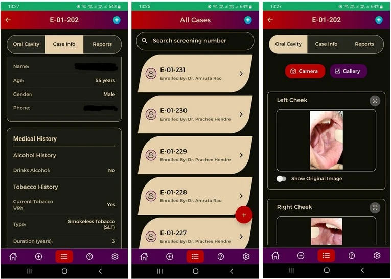
Finally, the trained model was deployed into a user-friendly app, with a backend and user interface. Using the latest industry-standard technology, the backend of the app was built to include a meta-data database, image database, and annotation database. The databases interact with each other ensuring no misalignment, with access to the data and security features in place.
The user-interface starts with the sequential appearance of a login feature, different access channels for different user profiles, and a consent form. In-app tutorials and explanations also guide the trained healthcare professional with best practices for image acquisition and subsequent processing steps. The app includes privacy settings that do not allow screenshots or recordings (so as to protect medical data) and has been extensively tested with oral experts and dentists, a process that is still ongoing. Following image capture and upload, the model scans the images and provides an analysis – in the form of a screening report – in a few seconds. In doing so, the AI-powered app – known as Mukhia Plus – provides oral cancer screening at one’s finger tips (3).
With the ISO 13485 certification and CDSCO Class B license (as software as a medical device) in place, Mukhia Plus is open to partnering with hospitals, clinics, insurance companies, as well as employee wellness and screening packages. The technology is being used on-site in corporate screening sites and dental hospitals by trained healthcare professionals. ‘Oral cancer screening has never been more non-invasive, accessible, and affordable’, says Ritwik. ‘With Mukhia Plus, all you need is a smart phone, internet connection, and the downloaded app’, he adds.
Beyond the development of a screening tool, the technology behind Mukhia Plus has also allowed Ritwik and his team to explore some blue-sky research in the field.
Ensuring the capture of high-quality images is an important aspect of image analysis research. In partnership with IIT Bombay, Edgescan AI is using algorithm-based methods to improve image collection, and therefore the raw images, that go into a model (4). ‘We have developed and incorporated a small intelligence system into the image capture feature – one that can flag poor images, ensure clarity and zoom, and point out shadows and other artefacts – specifically for oral cavity images’, states Ritwik. The work is soon to be presented in the 28th International Conference on Pattern Recognition in Lyon, France (4).
Another open research area in the field is training a model to perform well when the image data is not well-distributed. Explains Ritwik, ‘What we found was that while we had a large dataset of 62 different types of lesions, all of them did not have the same number of images. There were many images of normal oral cavities, and as expected, much fewer images of lesions identified as squamous cell cancers or salivary gland tumors’. AI-based models are very sensitive to skewed distribution of images, an effect known as the ‘tail effect’. The solution lies in developing methods to specifically improve model performance in these infrequent classes. ‘Long-tailed problems are well-known in healthcare data. Working with the team at Edgescan AI, we came up with a novel loss function to overcome the problems with long-tailed data sets’, says Dr. Kshitij Jadhav, a collaborator from IIT Bombay. ‘We titled our paper, Taming the Tail!’ adds Ritwik (5).
While these new advances might not directly be applied to the current version of the Mukhia Plus model, it allows the team at Edgescan AI to ‘broaden the horizons’ of this technological development. Says Ritwik, ‘It does create a pipeline of new research that can in the future be modified and plugged into new applications and tools. This is particularly relevant for the development of modifications that can overcome other limitations of AI based screening tools, such as algorithmic bias in image analysis and misidentification of lesions’.
With AI-based solutions rapidly transforming healthcare, Edgescan AI is a step forward for ‘augmented intelligence’ that could not only provide a tractable solution to a current public health problem but also contribute to future scalable technologies.
To know more about how you can engage with Mukhia+, write to ask@edgescan.ai.
Report written by Karishma S Kaushik, MBBS, MD, PhD
References
- https://www.pib.gov.in/PressReleasePage.aspx?PRID=2019532®=48&lang=2
- https://journals.lww.com/indianjcancer/fulltext/2010/47001/tobacco_and_health_in_india.3.aspx
- https://edgescan.ai/
- https://icpr2026.org/
- https://arxiv.org/abs/2410.04084
AI in Healthcare
Mukhia Plus: AI-Powered App Transforming Early Oral Cancer Detection in India